
We attribute the differences between charges in both data sets to different data collection and refinement schemes, different temperatures and a different accuracy of integration.įragment Bader charges ( ) for molecular species present in the crystal lattice (iii) formal +1 and −1 charges are well localized on the NH 4 + cation and oxalate anion, respectively.

(ii) oxalic acid molecules are either slightly negative or can be considered neutral General conclusions are basically the same in both cases, that is: The integrated fragment charges of all molecular species are summarized in Table 2. It is valuable to compare the modelled electron density distributions on the basis of the Bader quantum theory of atoms in molecules (QTAIM) analysis performed in both cases.

However, higher mosaicity may also be attributed to some extent to the size of a crystal and a higher number of imperfections, due to different synthesis procedures. It can also be noted that at higher temperature the extinction correction was not necessary, which may result from the increased mosaicity of a crystal. The use of Gram–Charlier parameters (Johnson, 1969 Kuhs, 1983 Scheringer, 1985 ) in the charge density distribution refinement is therefore justified. The latter shows that the effect observed on O10 at 100 K can be indeed attributed to the anharmonic thermal motion rather than disorder or other effects. (ii) at lower temperature anharmonic corrections for one of the O atoms (namely O10 in our original paper) are not necessary. (i) the unit cell is larger at 100 K than at 15 K Therefore, as expected, the following observations can be made: This data collection temperature constitutes a significant difference compared to our study conducted at 100 K. (2013 ) described the electron density of ammonium tetraoxalate dihydrate obtained at 15 K. §Values given for 10 121 reflections with. ‡Values given for all data (14 225 reflections). †Definition of the R mrg factor is summarized in the supporting information to our original article (Jarzembska et al., 2014 ). Selected parameters characterizing the X-ray data collection in both cases Selected parameters of the two data collections are summarized in Table 1. Such a comparison is especially valuable since the data had been measured using a well established methodology applying a Bruker AXS CCD-type APEX II detector at a synchrotron beamline known for accurate and precise electron density studies. Consequently, in this short addendum we would like to supplement our previous work by comparing the two charge density distributions. (2013 ) deals with the charge density distribution of the same compound, but measured at the 15-ID ChemMatCARS beamline of the Advanced Photon Source synchrotron (Chen et al., 2014 ). The corresponding CIF was not present in the standalone version of the Cambridge Structural Database (Allen, 2002 ) available at the time we published our contribution. Unfortunately, it transpires that we have overlooked a very important reference, namely the article published by Stash et al. A crystal of ammonium tetraoxalate dihydrate had been chosen as a test case due to its structural complexity, low symmetry and presence of anharmonic motion.

The facility also offers departmental clients access to the Cambridge Structural Database of well over 800,000 organic and organometallic structures.Very recently we published a paper describing the first use of a Bruker AXS CMOS-type PHOTON 100 detector in experimental electron density studies (Jarzembska et al., 2014 ). Some research groups with their own crystallographic computing resources use data collected by the facility to solve and refine their own structures. The crystallographer is available for consultation when structural material is to be published, and will gladly assist in the preparation of publication-ready material (tables, descriptions, diagrams, Crystallographic Information Files ) as required by the journal of interest.
#BRUKER APEX II CCD DETECTOR TEST FULL#
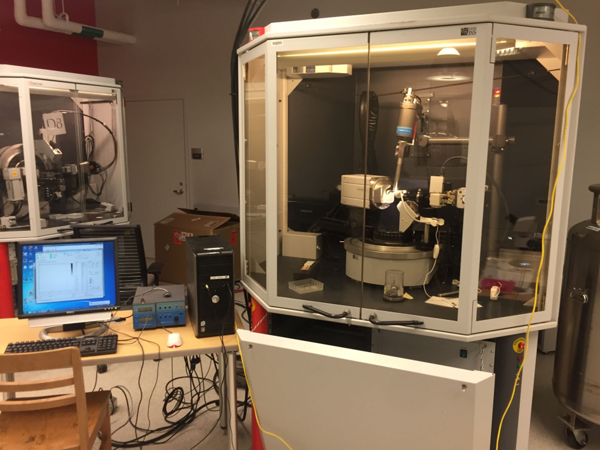
Structures are normally fully solved and refined by the staff crystallographer, with full structure reports (tables of experimental parameters, atomic coordinates, interatomic distances and angles, and structural diagrams) being supplied to the researcher(s). Crystal samples are usually cooled under a cold nitrogen stream during data collection in order to enhance data quality and to protect sensitive samples from decomposition. Data are collected using (a) a Bruker D8 Duo diffractometer / SMART APEX II CCD area detector system equipped with two radiation sources (Cu microsource and and Mo sealed-tube) or (b) a Bruker PLATFORM diffractometer equipped with a SMART APEX II CCD area detector and a sealed-tube (Mo) source. The X-ray Crystallography Laboratory (XCL) provides single-crystal X-ray crystallographic services to the Department.


 0 kommentar(er)
